Day :
- Reconstructive Surgery | Mohs Surgery | Hand Surgery | Body Lift
Session Introduction
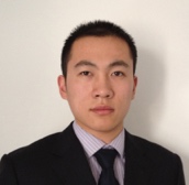
Fanjun Meng
Shandong Provincial Hospital, China
Title: Modified subcutaneous buried horizontal mattress suture compared with vertical buried mattress suture

Biography:
Fanjun Meng has his expertise in plastic and reconstructive surgery. The Modified Subcutaneous Buried Horizontal Mattress Suture he proposed in the paper is a new technique to close the tensioned wound. In vitro study and clinic practice, it is proved to be an efficient technique to reduce the tension of the wound and to prevent scarring postoperation of the large skin lesion excision.
Abstract:
Background: Wound tension reduction is still a challenge to surgeons. Over the years, many techniqueshave been proposed to avoid this issue. In this paper, we present a new suture technique.
Objectives: To investigate the tension-reduction effectiveness of the modified subcutaneous buried horizontal mattress suture compared with the vertical buried mattress suture technique.
Methods: Two suture techniques, the vertical buried mattress suture (group A) and the modified subcutaneous buried horizontal mattress suture (group B), were performed on paired samples of symmetrical skin flaps. An equal pulling force was applied to each paired sutured flap, and the dehiscences of the samples in the two groups were compared. Then, after the periodic mechanical pulling force was recorded, the dehiscences were compared again.
Results: The dehiscences of the vertical buried mattress suture samples(group A) were much wider than their corresponding samples. Modified subcutaneous buried horizontal mattress suture samples (group B) remained well closed with no or minimal dehiscence, under various
Lai Chih-Sheng
Taichung Veterans General Hospital, Taiwan, R.O.C
Title: Functional outcome and complications of robot-assisted free flap oropharyngeal reconstruction
Biography:
He is an attending plastic surgeon at Taichung Veterans General Hospital, Taiwan. He has more than 10 years of experience as plastic surgeon and specializes in wound treatment and reconstructive surgery. He obtained medical degree from Chung Shan Medical University. He has also authored many research publications and is an active member of many surgery societies as Taiwan Society of Plastic Surgery, Member of Taiwan Society for Surgery of the Hand, Member of Taiwan Society for Reconstructive Microsurgery
Abstract:
Purpose: The purpose of this study was to assess the outcomes of robotic-assisted oropharyngeal reconstruction comparison with conventional free flap reconstruction. The robotic surgical system provides a clear, magnified, 3-dimensional (3D) view as well as a precise and stable instrumental movement, which minimizes many technical difficulties that may be encountered in the surgical treatment of oropharyngeal tumors.
Materials and Methods:A retrospective review of consecutive patients who underwent reconstructive operations using free radial forearm fasciocutaneous flap for oropharyngeal defects over a 20-month period (May 2013 to December 2014). The primary predictor variable was method of reconstruction (conventional versus robot-assisted). Outcome measures were postoperative complication rates, revision rates, and postoperative functional outcomes.
Results:The study sample consisted of 47 subjects who underwent reconstructive operations using free radial forearm fasciocutaneous flap for oropharyngeal defects (33 conventional and 14 robot-assisted reconstructions). Complication rates between the conventional and robot-assisted groups were similar for flap failure, partial necrosis, wound infections, hematoma or seroma formation, wound dehiscence, and fistula formatiom. The revision requiring additional operation was comparable between the two cohorts. The functional outcomes postoperatively of robot-assisted reconstructions are better than conventional reconstructions as demonstrated by the Functional Intraoral Glasgow Scale scores.
Conclusion: There is no significant difference in complication and revision rates between conventional versus robot-assisted oropharyngeal reconstructions. The application of a robotic surgical system seems to be a safe option with better oral function postoperatively in the free flap reconstruction of oropharyngeal defects without lip or mandible splitting.
Chao Chen
Shandong University, China
Title: Microsurgical flow-through flaps for reconstruction of volar tissue defect of fingers

Biography:
Dr. Chao Chen is an attending doctor in Department of Hand and Foot Surgery of Shandong Provincial Hospital affiliated to Shandong University. He graduated from Shandong University, School of Medicine and got master degree of orthopedic in 2012. He got PhD degree of Clinical Anatomy in Southern Medical University in 2015, and his major research subject during PhD’s study is microsurgical anatomy, which dramatically improve his level of microsurgery.
Dr. Chen has been a microsurgeon since he finished orthopedic training in 2014. His department is one of the most famous microsurgical center in China, and he got good microsurgical training with guide of Professor Zengtao Wang and Dr. Liwen Hao. His specialties including hand surgery, limb replantation, thumb and finger esthetic reconstruction, vascularized tissue transplantation. He can successfully anastomose small vessel with caliber of 0.2mm. He has performed over 150 super microsurgeries (including fingertip replantation and mini-flap transplantation) and has total success rate more than 90%. Of all his more than 100 vascularized tissue transplantation surgeries, only one case failed.
Dr. Chen has intense study in microvascular anatomy associated with mini flaps in hand and foot. He has extensive experience in the mini perforator flaps harvested from hand and foot, especially for primary reconstruction of composite tissue defect in finger. He has published 4 SCI articles (coauthor in 3 articles) associated with microsurgery.
Abstract:
Backgrouds: Composite tissue defect of the volar surfaces of fingers are frequently associated with digital vessel damage. Different reconstructive methods were used for such injuries, like digital artery flap from adjacent finger, A-A typed flow-through venous flap, or vein graft combined with a regional flap. Flow-through glabrous flaps can provide esthetic tissue coverage as well as revascularization.
Methods: Between June 2010 and April 2017, we prospectively studied the use of Microsurgical flow-through glabrous flaps to achieve simultaneously digital revascularization and soft tissue coverage in 20 fingers of 17 patients who experienced volar injuries, comprising 7 great toe fibular flaps, 4 medial plantar flaps, 2 pedismedialis flap, 3 hypothenar flaps and 4 thenar flaps. The nerve passing through the great toe fibular flap or medial plantar flap was used to repair digital nerve defects.
Results: All flaps survived completely. During a mean follow-up period of 13.6 months, the majority recovered excellent appearance and function. The flaps had the characteristics of normal finger volar skin: hairless, with similar texture and color. The sensation of finger pulp which repaired with neurovascular flap gained satiscactory recovery.
Conclusions: Glabrous flow-through flaps provide excellent reconstruction for fingers with volar injuries associated with digital vessel damage. The great toe fibular flap and the medial plantar flap are reliable and useful options for complicated finger injuries associated with digital vessel and nerve injuries.Flow-through thenar flap is our first choice if the patient denied to harvest flap from foot.
Adilson Farrapeira Jr
Sobradinho Hospital Plastic Surgery Service, Brazil
Title: Global face management
Biography:
Adilson Farrapeira Jr has completed his plastic surgery specialization in 2008 at the Ivo Pitanguy Institute. Currently works as director of Adilson Farrapeira Plastic Surgery Institute, regent of Armed Forces Hospital Plastic Surgery Service and coordinator of Sobradinho Hospital Plastic Surgery Service.
Abstract:
Facial treatment is one of the greatest challenges in the plastic surgeon practice. A wide variety of procedures are available to improve skin appearance volumetric reposition and tissues tightening. To obtain a natural result is necessary a facial evaluate in its three dimensions, acting on all layers of tissue, from the skeletal framework to the epidermis. The facelift surgery continues being the gold standard for facial rejuvenation, bringing the best results. However the new technologies, have great importance to complement the surgery result. This study aims to demonstrate the experience of the senior author in the global face management, associating video endoscopic surgery, several techniques for the middle and lower face treatment and ancillary procedures such as lasers, botulinum toxin and fillers.
Pusit Jittilaongwong
Punisa lip and plastic surgery clinic, Thailand
Title: Experience of 7145 cases on Lip reduction surgery in Thailand

Biography:
Dr.Pusit, the founder of Punisa lip and plastic surgery clinic. He is a Broad certified plastic surgeon graduated from Surgery Department, Siriraj Hospital Medical. Dr Pusit’s name is one of the most sought-after plastic surgeons for lip surgery. He is also well known as the pioneer lip reduction “Krachap lip” surgery in Bangkok, Thailand.He was invited to speak in conferences 27 th Annual meeting of The Society of Aesthetic Plastic Surgeons of Thailand 2017.He also contributed to many articles published in newspapers as well as appeared in social media.Dr. Pusit has worked in private practice actively improving the appearance of patient’s lips by reducing their size and abnormality for thousands of patients for the past 7 years
Abstract:
The purpose of this report is to present my personal experiences over the last 7 years in lip reduction surgery.
A method of evaluating the results of lip reduction surgery was performed on 7145 consecutive patients from 1 January 2011 to 30 November 2017 at Punisa Clinic lip surgery clinic. Patients who have undergone a lip reduction surgery by Dr. Pusit technique, “Seagull wing incision” can expect more desired shape & size of their lips and even an improved smile, abnormality. Results of my patients included 5156 cases underwent upper lip surgery, 291 cases for lower lip surgery and 1698 cases for both upper-lower lip surgeries. Patients who underwent lip reduction surgery reported an overall of high satisfaction rate with their surgical outcome. In conclusion, Lip reduction surgery tend to be more popular in Asian countries from past until now. Lip surgery can improve patients lip shape, size and smile. The minor complication were reported with asymmetry, scarring (Keloid) and lip tightness
- Mohs Surgery| Cosmetic Dermatology| Breast Surgery | Burn Care/ Trauma Surgery
Session Introduction
Paulo Renato de Paula
Federal University of Goias - Brazil
Title: Experience of ten year’s with a conically shaped implant: Breaking the paradigm

Biography:
Paulo Renato de Paula has completed his Plastic Surgery training in 1995 from Prof. Pitanguy’s plastic Surgery Program; his M.Sc. at the age of 35 years from Federal University of Rio de Janeiro and his PhD at the age of 51 years from Federal University of Goias. He is an Adjunct Professor and Head Chief of Plastic Surgery Unit at School of Medicine–Federal University of Goias. He is a Supervisor of Residency Program and Internship in Plastic Surgery of the University. He has 12 book’s Chapter (including International book as author and co-author), 12 papers as an author and co-author and 32 studies in meetings as author and co-author (presented or e-poster).
Abstract:
Introduction: Breast implants are often used for the reconstructive and cosmetic purpose, for pure augmentation or associated with mastopexy, demonstrating low morbidity and a reduced number of complications. These procedures demonstrated a significant improvement in the quality of life, like individual/social well-being, self-confidence, and favorable psychological consequences. The innumerable options of breast implants and their variants in the market allow us to offer more specific results for each of breast/thorax and patient wish, with a high degree of satisfaction. The most common implants used are the round or anatomical shape.
Objective: The present study aims to demonstrate a conical breast shape implant. A device model with different angle, shape, and projection and can be a great option.
Method: It is a descriptive and retrospective 10 years’ study with the use of breast implants with a conical shape, then use it and a study with patients’ satisfaction’s degree with these models.
Results: A total of 1182 implants (591 patients) were used during the study period, of which 552 implants (276 patients) were the conical shape (46,7%), all with polyurethane coating, in pre-pectoral (retroglandular/retrofascial) location in 92,2%. Inframammary access was used in 84.6%. The mean volume was 250,65 and mean age was 32 years. The follow-up time ranged from 6 to 120 months, with an average of 78.5 months. Small complications occurred in 3% (small dehiscence, asymmetry, aestrias, hypertrophic scar). Only two contractures (unilateral) cases after 5 years and no extrusion happened. A questionnaire was carried out to evaluate the degree of satisfaction. 85.3% responded and of these, 96.5% declared themselves very satisfied and satisfied with the implant profile and 3.5% were not satisfied. There was no case of dissatisfaction.
Conclusion: Cone-shaped implants are an excellent option in the surgical arsenal of breast implants according to the patient's desire and correct indication, with few complications and a high degree of satisfaction.
Lin-Gwei Wei
Taipei Medical University, Taipei, Taiwan, ROC
Title: 500-gray γ-irradiation may increase adhesion strength of lyophilized cadaveric split-thickness skin graft to wound bed
Biography:
Trained by the National Defense Medical Center of Taiwan, Lin-Gwei Wei is the only plastic surgeon in a countryside, 690-bed, military, teaching hospital, while he also works part-time in another medical center with his mentor Prof. Hsian-Jenn Wang. The busy schedules of surgeries do not suppress Dr. Wei’s curiosity about mysteries of human body, and he tries to work out some difficult clinical challenges with researches. Currently, he participates in the complex “bank of artificial skin” development program of Prof. Wang, in a clinical trial of wound-healing enhancing gel, and in an electromyographically-controlled prosthetic limb development program. He hopes that his wide interests in burn care, acute and chronic wound care, trauma care, local- and free-flap surgeries can eventually do some good to researchers and to the people, just like what he has done in his clinical practice.
Abstract:
Background: Human cadaveric skin grafts are considered as the “gold standard” for temporary wound coverage because they provide a more conductive environment for natural wound healing. Lyophilization, packing, and terminal sterilization with gamma-ray can facilitate the application of cadaveric split-thickness skin grafts, but may alter the adhesion properties of the grafts. In a pilot study, we found that 500 gray (Gy) gamma-irradiation (γ-irradiation) seemed not to reduce the adherence between the grafts and wound beds.
Aim and Objectives: We conducted this experiment to compare the adherences of lyophilized, 500-Gy-γ-irradiated skin grafts to that of lyophilized, non-irradiated grafts.
Materials and Methods: Pairs of wounds were created over the backs of 12 Sprague Dawley rats. Pairs of “lyophilized, 500-Gy-γ-irradiated” and “lyophilized, non-irradiated” cadaveric split-thickness skin grafts were fixed to the wound beds. Adhesion strength between the grafts and the wound beds were measured and compared.
Results: On post-skin-graft day 7 and day 10, the adhesion strength of γ-irradiated grafts was greater than that of the non-irradiated grafts.
Conclusion: Because lyophilized cadaveric skin grafts can be vascularized and the collagen of its dermal component can be remodeled after grafting, the superior adhesion strength of 500-Gy-γ-irradiated grafts can be explained by the collagen changes from irradiation
Laura Bamford
York Teaching Hospital, UK
Title: Reconstruction of a periorbital defect with Radial Forearm Free Flap following necrotising fasciitis
Biography:
I graduated from the University of Leeds, UK, with BChD in Dental Surgery. I am currently enjoying my second year as a Senior House Officer (or Core Trainee 2) in the Oral and Maxillofacial Surgery unit at York Teaching Hospital NHS FT. I am deputy editor of the Patient Safety Matters Staff Bulletin for the hospital. Prior to this I worked briefly as a Work Psychologist as I hold both a BSc and MSc in Psychology. It is my intention to compete a third SHO year before returning to university to study medicine, to embark on a career as a Maxillofacial surgeon
Abstract:
Introduction: A 43-year-old male presented with a 2-day history of increased pain and swelling around his right periorbital region following a small abrasion to his eyebrow. On presentation, necrotizing fasciitis was clinically diagnosed. Two-stage debridement involved the extensive sacrifice of extensive soft tissues including orbicularis oculi and levator muscles and eyelids, the globe was spared.
Reconstruction: The reconstructive challenge included separate coverage of the eyelid and minimizing bulk to the surrounding periorbital region. This case was jointly managed with the Oculoplastic Surgeons. Previously documented reconstruction with myofascial free flaps has led to unwieldy flaps with aesthetically poor results. To maximize the aesthetic result skin grafts from the upper arm were grafted to the eyelids and were then completely covered with a 10x7cm soft tissue radial forearm free flap (RFFF) utilizing cephalic venous drainage. This was anastomosed to the facial vessels. Secondary surgery was performed 8 weeks later involving division of the flap, uncovering the skin graft and debulking to provide contour.
Conclusion: In this unusual case, composite reconstructive approaches were combined to overcome a unique challenge. This is the first described case of using RFFF for reconstruction of the periorbital region following such extensive tissue loss, whilst maintaining the function of the eye following necrotizing fasciitis. The RFFF provided excellent short and long-term reconstruction. It protected the eyelid skin grafts and matched the facial contours well. Division of the flap following the establishment of the collateral blood supply was straightforward and well tolerated. We would recommend its consideration for facial defect consideration once the acute infection is cleared.
Yair Shachar
Hadassah University Medical Center, Israel
Title: Expanded bi-pedicled “sleeve†flap for reconstruction of the upper extremity after large circumferential nevus excision in children
Biography:
Dr. Shachar is the chief resident at Hadassah Medical center plastic and reconstructive surgery. Working under the supervision of Prof. Alexander Margulis, the department has become a reference center in Israel for pediatric plastic surgery. Our chief expertise is pediatric tissue expansion for congenital and acquired deformities. As such, we perform more than a hundred cases of reconstructions for congenital melanocytic nevi annually.
Abstract:
Background: Large congenital melanocytic nevi entail significant medical and cosmetic ramifications for patients and families. Reconstruction is a challenging endeavor, especially when the lesion is present on the limbs. The literature describes various methods by which a plastic surgeon can address reconstruction; yet, to date, there has been no series describing a method that provides consistent results with low complication rates.In this study, we describe our surgical technique for reconstruction of the upper extremity after excision of large circumferential cnm with a pre-expanded bi-pedicled flap, namely the “sleeve” flap.
Methods: a systematic review of our database of pediatric patients treated for large and giant nevi was performed. Patients with large and giant circumferential upper extremity nevi were retrieved, and their charts reviewed for demographics, number of procedures performed, duration of follow-up, and complications.
Results: over a course of 12 years, eight patients with large or giant circumferential nevi of the upper extremity were treated at our institution with “sleeve” flap reconstruction. Mean follow-up time was 36 months. A single complication was seen. All reconstructions achieved satisfactory results, both functionally and cosmetically discussion: we describe our surgical approach for treating upper extremity large and giant circumferential nevi with pre-expanded bi-pedicled “sleeve” flaps. When properly planned and executed, this technique enables successful treatment of large and giant nevi of the arm and the forearm. Although arduous and complex, the process yields excellent aesthetic results with low complication rates. This technique is promising as the reconstructive option of choice for these difficult lesions.
- Facial Surgery | Rhinoplasty| Cosmetology & Skin Care
Session Introduction
Sherine Raveendran
Toronto Medical Aesthetics, Canada
Title: Classification and morphological variation of the frontalis muscle and implications on the clinical practice

Biography:
Ms. Raveendran is a Plastic, Reconstructive and Aesthetic surgeon with a special interest in burns and clefts. She was board certified as a specialist in Sri Lanka, continued her training in Plastic & Reconstructive surgery in the United Kingdom, and was successful in the Fellowship examination by the Royal College of Surgeons of Edinburgh and the European Board of Plastic surgical examination. She worked in many of the prestigious hospitals in the United Kingdom. She received her Masters degree in Aesthetic Surgery from Queen Mary University, London and was trained in cosmetic surgery in the UK.
Ms. Raveendran had an excellent academic record and won awards and medals during her training. She has published many scientific articles and authored book chapters in the field of Plastic and Aesthetic surgery. She has presented at several international meetings and is an invited reviewer for scientific journals. After practicing in the surgical field for more than a decade, Ms. Raveendran established Toronto Medical Aesthetics, an institute that provides advanced medical aesthetic procedures in the province of Ontario.
Abstract:
Forehead aesthetics is an imperative element in the rejuvenation process of the face. Restoration of the upper facial aesthetics facilitates to counteract the changes related to the aging process. The interaction between the frontalis and its antagonist's muscles contribute to the overall aesthetic balance of the forehead. In this study, we evaluated the gross anatomy of the frontalis and classified the muscle according to the morphological appearance. Twenty-six cadavers of Caucasian and South East Asian origin were dissected. The frontalis muscle was dissected without mobilization and the gross anatomy and variations were analyzed on the backdrop of gender and ethnicity. Our dissection studies revealed three main variations of the muscle based on the extent of interdigitation between the two bellies in the midline. The average length of the muscle is 10.9 cm in males and 9.1cm in females. The width of the muscle in females was 6.5 cm at the base and 4 cm at the insertion and 5.8cm at the base and 4.4cm at the insertion in males. In six specimens, the muscle attached only up to the medial two-thirds of the eyebrows. This was predominantly observed in the Caucasians cadavers and may contribute the constitutional downward slanting eyebrows in some individuals. This study provides a comprehensive analysis and classification of the frontalis muscle. Understanding the morphological variation of the muscle helps to amend the clinical application accordingly. Clinical evaluation of the patterns of decussation of the frontalis muscle may assist with non-surgical interventions using botulinum toxin.
Leila Freire Rego Lima
Jurado's Institute of Education and Research, Brazil
Title: Update in alar base reduction in rhinoplasty
Biography:
Leila Freire Rego Lima, ENT doctor, Facial Plastic Surgeon, Chief preceptor of the Jurado Institute of Post graduation in Facial Plastic Surgery-São Paulo/Brazil.
Abstract:
Purpose of review: The purpose of this article is to further explore the techniques available for alar base reduction by a recent review in this topic, focusing in the current advances in the field, in a clear and readable format.
Recent findings: Among the numerous techniques available, the management of cases with wide nasal base and alar flaring remains limited to three options: cinching sutures to pull in the alae together; or to use alar base excisions that remove tissue from the alar lobule to decrease flare and from inside the nostril to decrease width; and flap advancement.
Summary: Surgical modification of the nasal base is not a routine part of rhinoplasty and should be performed in a conservative manner to prevent complications like nasal stenosis. Moreover, this maneuver should always be the last one in rhinoplasty, as alar base modifications cannot be properly assessed until all other steps have been carried out.
Guillermina RamÃrez Orozco
Benemérita Universidad Autónoma de Puebla, Mexico
Title: Strategies for a successful rhinoplasty

Biography:
Guillermina Ramirez Orozco has her expertise and experience in complete evaluation of the nose; and bases her medical practice taking a holistic view of the patient. She sees the nose not only as a part of the respiratory tract, but also as the center of the facial beauty; her surgical techniques ensure the patient adequate respiratory function in addition to seek a permanent aesthetic harmony. Never sacrifices the function by the beauty. Guillermina has been professor of Otolaryngology and Rhinology in Benemérita Universidad Autónoma de Puebla and in the Universidad Popular Autónoma de Puebla for several years. Has worked for public and private institutions for over 15 years; and has collaborated with the “Manual para el Examen Nacional de Residencias Médicas” a book edited by Benemérita Universidad Autónoma de Puebla. Her interests include teaching, Neurolinguistic Programming, emotional intelligence, as well as accelerated learning techniques for all ages
Abstract:
Rhinoplasty is one of the most common procedures in facial surgery, and due to the delicate and complicated structure of the nose, it is also among the most difficult to carry out successfully. It involves a precise balance between shaping cartilage, bone, and soft tissue. However, those are not the only factors that affect the final result and even the convalescence of a patient.
The surgical team organization, anesthesia and the methods used to handle all the tissues involved during the procedure are critical to the outcome. Post-surgery, the healing and tissue contracture may persist for up to a year (some authors would even say two years).
Since the nose is the most prominent feature of the face, the skills and experience of the surgeon are extremely important in achieving success. Both the novice surgeon as well as the more experienced one, must take care of key aspects of the surgery to successfully achieve both the functional and aesthetic objectives for the patient.
Each case has its own challenges and requires a careful evaluation of the deformity preoperatively, and a clear understanding of the techniques available and most appropriate for correction. Every surgical operation is prone to complications. The only way to avoid the risk of such complications would be to not operate in the first place. A successful surgery requires a sequenced plan of action, together with a meticulous, uncompromising execution of the whole surgical event that gives protection to the surgical event.
